Grapevine disease pictures: what you're actually looking at
TL;DR
- The grapevine diseases you'll photograph most often are powdery mildew, downy mildew, Botrytis bunch rot, black rot, Eutypa dieback, Phomopsis cane and leaf spot, crown gall, Pierce's disease, fanleaf degeneration, and bunch stem necrosis.
- Each has a distinct visual signature.
- Get the ID right before you spray.
- It saves money and keeps your WPS records defensible.
Why do grapevine disease pictures actually matter for diagnosis?
A photo is not a diagnosis. Say that to yourself before you share a blurry image in a Facebook group and act on the replies. But a sharp, well-lit photo taken at the right growth stage, paired with your spray record and weather log, is the fastest path to a correct ID, especially when your farm advisor or extension agent is two counties away.
Visual diagnosis has real limits. Powdery mildew and downy mildew can look almost identical on a leaf underside if the light is wrong. Eutypa dieback and Esca produce similar shoot stunting early in the season. A picture helps only when you know which feature separates one disease from another, and you know what part of the plant to photograph.
This guide describes what to look for in each major disease photo, what you should capture in the frame, and where each disease sits in the season. It also tells you when a photo alone falls short and you need a lab test or PCR confirmation.
What does powdery mildew look like on grapevines?
Powdery mildew, caused by the fungus Erysiphe necator (formerly Uncinula necator), is the most photographed grapevine disease in the world. Good reason for that. It's the most economically damaging fungal disease in most wine regions [1].
On leaves, you'll see a white to gray powdery coating, usually starting on the upper surface but visible on both sides. Young leaves curl or pucker. In the field, hold the leaf at a low angle to sunlight: the mycelium catches the light and you can see it clearly before a phone camera picks it up. When you photograph it, shoot in diffuse light, not direct sun, and frame the whole leaf plus one close crop of the colony margin.
On clusters, infected berries develop a white coating at or just after fruit set. As berries grow, the fungus colonizes the skin, which then splits or cracks and becomes a gateway for Botrytis. Berries infected before 10-15mm diameter often show a net-like russeting scar at harvest. That scar is a common photo ID question from winemakers who missed the bloom-time infection.
In wood, look for the brown-to-purple blotching on green shoots, sometimes called "olive oil" patches. These are easy to miss and easy to confuse with Phomopsis lesions. The key difference: powdery mildew lesions on shoots are more diffuse, without the defined dark margins that Phomopsis produces.
UC IPM's grape powdery mildew guidelines use the term "web-like mycelium" to describe early colony appearance [1]. That's exactly what to look for in your photo: a spider-web texture before it thickens into the familiar white felt.
How do I tell downy mildew apart from powdery mildew in photos?
This is the single most common misidentification in the field, and it costs vineyards real money in wrong-product applications.
Downy mildew (Plasmopara viticola) shows up on the upper leaf surface as yellow-green, oily-looking spots called "oil spots." They're translucent when you hold the leaf to the light. Flip the leaf over and you'll find a white, cottony sporulation on the underside directly below each spot. That underside fuzz is the diagnostic feature. Powdery mildew produces its white coating on the upper surface. If the fuzz is on the bottom, it's downy [2].
Downy mildew needs liquid water and warm temperatures (59-77°F optimal) to release spores, so symptoms explode after rain events. Powdery mildew spreads in dry, warm conditions and is suppressed by rain. Your recent weather log is as useful as the photo.
On clusters, downy mildew causes what some texts call "gray rot of clusters," but the more diagnostic picture is the browning and shriveling of young berries and rachises after a sporulation event. The berries may turn brown and mummify, which looks similar to bunch stem necrosis and to late-season Botrytis. Take a close shot of the pedicel and rachis surface, looking for white sporulation on the stem tissue itself.
The Cornell University viticulture program and Washington State University extension both maintain photo libraries showing these distinctions [3][4]. If you're building a reference folder for your crew, those are worth bookmarking and printing.
What do Botrytis bunch rot photos show, and when does it spread?
Botrytis cinerea is a soft-rotting fungus that most growers know by feel before they see it clearly: affected berries are soft, and when you squeeze them the skin slips. In photos, the gray, dusty sporulation is the tell. The "gray mold" name is accurate. That gray powder is masses of conidia.
Early infections on flowers look like small brown flecks, easy to miss. The explosion happens after veraison when berry sugar rises and skin integrity drops, especially after rain or heavy dew. In tight-clustered varieties, Botrytis moves berry to berry through direct contact.
In photos, the gray sporulation is often confused with other molds, including species of Aspergillus that appear in hot climates. The color and texture differ: Botrytis is gray-brown and dusty, Aspergillus niger is black and powdery. Both warrant attention, but only Botrytis is the target of most fungicide programs.
Botrytis can also cause a shoot and cane blight in spring, producing brown lesions at the base of shoots that can kill the shoot tip. Those spring images look nothing like bunch rot and are often under-photographed and under-reported.
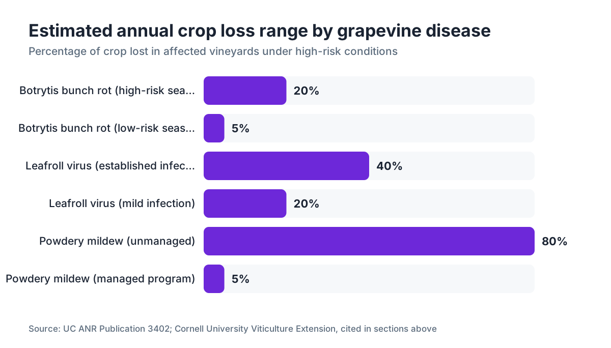
Botrytis bunch rot causes estimated losses of 5 to 20 percent of crop annually in high-risk regions, according to UC ANR Publication 3402 [5]. The range is wide because the disease tracks the weather so closely.
What does black rot look like on grapevines, and where does it start?
Black rot (Guignardia bidwellii) is mostly an eastern US problem, common in Virginia, New York, Pennsylvania, and the mid-Atlantic, but it shows up wherever humid summers follow wet springs.
On leaves, start with the classic reddish-brown circular lesion with a yellow halo. Within a week or two, tiny black dots (pycnidia) appear in the lesion center, often in a ring pattern. Those pycnidia are diagnostic: use a hand lens in the field or a macro lens on your phone, and photograph them close. No other common grapevine leaf disease produces pycnidia in that arrangement.
On berries, black rot produces what growers call mummies: hard, black, shriveled berries that hang in the cluster or drop to the ground. A cluster with mummies at harvest is a red flag for next year because those mummies are the main overwintering inoculum source [6]. Photograph them in context, showing the whole cluster and then a close shot of one mummy.
Timing narrows the ID. Black rot infects from pre-bloom through about four to six weeks after bloom. If you're seeing new berry infections after that window, suspect another pathogen.
Cornell's Integrated Pest Management program has kept a detailed black rot photo guide online since the early 2000s, and it's updated regularly [3].
How do you identify Eutypa dieback versus Esca in the field?
These two wood diseases are the most photographed and most debated diseases in old-vine vineyards, and they get confused constantly. Both cause stunted, chlorotic shoots with small, distorted leaves. Both come in through pruning wounds. Both are ruinous in old vines. They aren't the same disease, and they don't have the same management options.
Eutypa dieback (Eutypa lata) shows up as a wedge-shaped canker in cross-section of the wood, usually straw-yellow to brown, clearly defined against healthy white wood. Cut a cane or spur back to a larger wood section and photograph the cross-section. The wedge shape, pointing toward the pith, is the signature. Shoots growing from infected wood are stunted, with small cupped leaves and poor cluster development.
Esca is a complex of wood-rotting fungi (Phaeoacremonium aleophilum, Phaeomoniella chlamydospora, and Fomitiporia mediterranea are the main players) and produces a different wood picture. Internally, you'll see a soft, white, spongy rot in the central wood. On leaves, you get the "tiger stripe" pattern: interveinal chlorosis with green veins surrounded by yellowing that turns brown. The tiger-stripe leaf pattern is the most-photographed Esca symptom, and it's hard to mistake once you've seen it [7].
Esca also produces an acute form called apoplexy, where an entire vine collapses and dries within days in summer. Photograph the whole vine and then a cross-section of the trunk.
Neither disease is curable once established. The management question is slowing the spread, and that starts with an accurate ID.
What does Phomopsis cane and leaf spot look like?
Phomopsis viticola is an early-season disease that infects shoots and leaves during the first few inches of growth in spring. The window is short: roughly from bud break to about 15cm shoot length, during wet, cool weather.
On shoots, look for small, dark brown to black lesions at the base of the first several internodes. These lesions have a bleached center with a dark margin and often leave the cane looking bleached or silvery on infected sections by harvest. That silver or bleached look on dormant canes in winter is one of the most useful photographic clues: a bleached basal internode with a dark dot pattern (pycnidia again) is almost always Phomopsis.
On leaves, the lesions are small (2-3mm), angular, yellow-margined spots that are easy to overlook. Severely infected leaves may show ragged holes as the dead tissue falls out. Photograph in early morning with backlit conditions: the yellow halo shows well.
On clusters, Phomopsis causes a bunch rot that resembles Botrytis but starts from the rachis inward. The affected rachis turns brown and the cluster shrivels. This is less common than the cane and leaf form but devastating when it hits.
WSU's extension viticulture resources cover Phomopsis in the Pacific Northwest, where wet springs make it a regular problem [4].
What does crown gall look like, and is a photo enough to confirm it?
Crown gall (Agrobacterium vitis) produces swollen, irregular, tumor-like growths on trunks, cordons, and canes, usually at wound sites, pruning cuts, or graft unions. In photos, young galls are soft and light-colored, almost white or cream, with a rough, warty surface. As they age and die back, they turn brown, corky, and crack. Large galls on old trunks can reach the size of a grapefruit.
A photo is usually enough for a confident field call in classic cases. The location (at or near the graft union or a wound), the irregular cauliflower-like texture, and the vine decline that comes with it are all pretty clear. Lab confirmation (PCR or culture) matters when you're making replanting decisions or buying nursery stock.
Frost cracks and similar wounds can produce callus tissue that looks gall-like to an untrained eye. The difference: callus is smoother and more uniform. Crown gall tissue is irregular and often has dead sections right next to actively growing tissue.
Crown gall is systemic. Once a vine is infected, the bacteria persist in the vascular system, which is why a single cold-injury winter can trigger widespread gall expression across an established vineyard [12].
How do you spot Pierce's disease symptoms in photos?
Pierce's disease (caused by the bacterium Xylella fastidiosa, spread by sharpshooter leafhoppers) has a very specific leaf symptom called island chlorosis, or leaf scorch. The margins of the leaf turn brown, but a narrow band of yellow separates the brown margin from the green interior. That yellow band is the diagnostic marker [8].
Another classic photo feature: matchstick petioles. Late in the season, the leaf blade drops off but the petiole stub stays attached to the cane. A cane covered in dry petiole stubs with no leaf blades is a strongly suggestive picture. Pair that with marginal leaf scorch and you have a compelling visual case.
Berries shrivel unevenly, with some berries in a cluster affected and others normal, producing what's called a raisin cluster. Internal wood symptoms require cutting: the wood shows green islands of tissue in cross-section.
Pierce's disease sits throughout California's Central Valley and Southern California and is spreading north. It's also present in the Gulf Coast states. In California, the Glassy-winged Sharpshooter became the primary vector in the late 1990s, causing heavy vineyard losses in Temecula. Photographs of GWSS and leaf scorch together make a solid case for your county ag commissioner.
Confirmation requires ELISA or PCR testing. Do not rely on photos alone when making removal decisions [8].
What do leafroll virus and fanleaf virus look like in photos?
Grapevine leafroll disease is caused by a complex of closteroviruses (GLRaV-1 through GLRaV-9, with GLRaV-3 being the most widespread and damaging). The symptoms differ between red and white varieties, which changes everything for photo diagnosis.
In red varieties, infected vines show brilliant red or purple interveinal coloration starting at veraison, with the leaf margins rolling downward. The leaf veins stay green, so you get a striking red leaf with green veins. That pattern is hard to miss from any distance. In white varieties, the color change is yellow or pale green with the same downward rolling and green vein pattern, much harder to see and often missed.
Fanleaf degeneration (caused by Grapevine fanleaf virus, GFLV) produces a very different picture. Leaves develop an irregular, fan-like shape with abnormal lobing, teeth, and asymmetry. Shoots may show zigzag growth and double nodes. Clusters are small, irregular, and sometimes show shot berries (unfertilized berries). The chlorotic form shows yellow mosaics on leaves [9].
Both leafroll and fanleaf are confirmed by ELISA or PCR testing. A photo diagnosis is suggestive but not definitive for either, especially for leafroll in white varieties or when symptoms are mild.
Grapevine leafroll disease can cut yields by 20 to 40 percent and delay ripening by 10 to 21 days, according to a frequently cited Cornell study [3].
What's the right way to photograph grapevine disease symptoms for a lab submission?
If you're sending photos to an extension specialist, your county farm advisor, or a plant diagnostic lab alongside a physical sample, the photos should tell a story, more than document a single lesion.
Shoot in this order. First, a wide shot of the whole vine or block showing the pattern of affected plants (randomly scattered vs. row-end vs. spreading from a single point tells you a lot about cause). Second, a mid-range shot of an affected shoot or cluster in context. Third, a close-up of the lesion, ideally with a coin or pen for scale. Fourth, if it's a wood disease, cut a section and photograph the cross-section fresh, within a minute of cutting, before oxidation changes the color.
Always note the date, the variety, the row and vine ID, and your recent weather and spray history in the same message or file. A photo without that context is far less useful to a diagnostician.
For physical samples, the UC Davis Plant Diagnostic Center and most state land-grant labs post submission guidelines online. Shipping live, moist plant material in a sealed plastic bag speeds up decay and degrades the sample. Use paper bags or damp paper towels inside a breathable package and ship Monday through Wednesday to dodge weekend delays in transit [10].
This is where spray recordkeeping earns its keep. When a specialist asks what you've sprayed in the last 30 days and at what rates, you need that answer fast and accurate. Tools like VitiScribe are built for that kind of field-to-lab communication, letting you pull your spray history and block notes from the same screen where you're documenting symptoms.
The EPA Worker Protection Standard requires pesticide application records to be kept for two years and made available to workers on request [11]. A disease ID event is exactly when those records prove their value.
Disease symptom comparison: what to look for at a glance
The table below summarizes the key visual features for the most common grapevine diseases. Use it as a field reference when you're looking at photos or standing in the row.
| Disease | Primary location | Key visual feature | Season |
|---|---|---|---|
| Powdery mildew | Leaves, shoots, clusters | White powdery coating, upper surface | Bloom through veraison |
| Downy mildew | Leaves, clusters | Yellow oil spots upper; white fuzz lower | Spring through summer |
| Botrytis bunch rot | Clusters | Gray dusty sporulation on berries | Veraison through harvest |
| Black rot | Leaves, berries | Circular lesions with pycnidia; berry mummies | Pre-bloom to 6 wks post-bloom |
| Eutypa dieback | Wood, shoots | Wedge-shaped canker in cross-section | Visible spring/summer |
| Esca | Wood, leaves | Tiger-stripe leaves; white spongy wood rot | Mid-summer |
| Phomopsis | Canes, leaves | Bleached basal internodes; pycnidia | Early spring |
| Crown gall | Trunk, cordons | Warty tumor at wound sites | Year-round |
| Pierce's disease | Leaves, berries | Marginal scorch with yellow band; matchstick petioles | Summer |
| Leafroll virus | Leaves | Red leaves with green veins; downward roll | Veraison onward |
| Fanleaf virus | Leaves, shoots | Fan-shaped leaves; double nodes | Spring through summer |
| Bunch stem necrosis | Cluster stems | Brown, shriveled rachis; no sporulation | Veraison |
Sources: UC IPM, Cornell Viticulture, WSU Extension [1][3][4][5]
When should you call a lab instead of relying on photos?
Photos are fine for screening and for flagging what to watch. They fall short in four situations.
First: when you're making a vineyard removal decision. Crown gall, Pierce's disease, leafroll, and Eutypa in old vineyards are expensive calls. Confirm with lab work before you pull vines.
Second: when you're seeing new symptoms you've never seen before on your property. A novel symptom pattern deserves a formal submission.
Third: when a fungicide program isn't working. If your mildew spray program is on schedule but disease keeps showing up, you might be dealing with fungicide resistance, or you might be misidentifying the pathogen. Lab work sorts out which.
Fourth: any time you suspect a regulated pathogen. Pierce's disease (Xylella fastidiosa), grapevine trunk diseases in some states, and certain virus diseases trigger notification requirements. Your county agricultural commissioner needs a lab-confirmed report, not a phone photo [8].
The UC Davis Plant Diagnostic Center and the Cornell Plant Disease Diagnostic Clinic are two of the most capable labs for grapevine diseases in the US [10][3]. Most state land-grant universities run their own diagnostic labs too. Turnaround is typically one to two weeks for standard testing; PCR confirmation can come back faster.
After results land, document them alongside your spray records, variety maps, and photos. If you're managing multiple blocks or dealing with an insurance claim, a clean, time-stamped record that links symptoms to confirmed diagnosis to treatment response is the only paper trail that holds up. VitiScribe's field log lets you attach photos and lab reports to individual block records, so that chain of evidence stays intact without a filing cabinet.
Frequently asked questions
What are the most common grapevine diseases to identify from photos?
Powdery mildew, downy mildew, Botrytis bunch rot, black rot, Eutypa dieback, Esca, Phomopsis, crown gall, Pierce's disease, leafroll virus, and fanleaf virus are the diseases extension specialists see most often in photo submissions. Powdery mildew and Botrytis are by far the most common and the most economically significant across most US wine regions.
Can I identify grapevine diseases just from a photo on my phone?
For textbook cases of common diseases like powdery mildew or crown gall, a clear photo is usually enough for a confident field call. For wood diseases, virus diseases, and anything you're seeing for the first time on your property, a photo is a starting point, not a conclusion. Lab confirmation is necessary before making vine removal or major spray program changes.
What's the difference between Eutypa dieback and Esca photos?
Eutypa shows a wedge-shaped canker in wood cross-section, with stunted shoots growing from infected spurs. Esca shows white spongy internal wood rot and, on leaves, a tiger-stripe pattern of interveinal yellowing with green veins. Both produce stunted, chlorotic shoots, which is why a wood cross-section photo is essential for telling them apart. Neither is curable once established.
What does powdery mildew look like on grape berries vs. leaves?
On leaves, powdery mildew shows as white-gray powdery coating on the upper surface, with young leaves puckering or curling. On berries, early infection causes the same white coating, but berries infected before 10-15mm often show only a russeting scar at harvest. Heavily infected young berries crack and become entry points for Botrytis.
How do I photograph grapevine disease symptoms for an extension specialist?
Take four shots: a wide view of the affected area in the block, a mid-range of the affected shoot or cluster, a close-up of the lesion with a coin for scale, and a cross-section photo if a wood disease is suspected. Include the date, variety, block ID, and recent spray history in your message. Diffuse light works better than direct sun for most leaf symptoms.
What does Pierce's disease look like, and how do I confirm it?
Pierce's disease causes leaf margin browning with a narrow yellow band separating the brown from the green interior. Late in the season, petiole stubs stay on the cane after leaf blades drop, called matchstick petioles. Confirmation requires ELISA or PCR lab testing. Don't make removal decisions on visual symptoms alone, and report confirmed cases to your county agricultural commissioner.
What are the tiger-stripe symptoms on grapevine leaves?
Tiger-stripe is the common name for the leaf symptom of Esca disease, one of the major trunk disease complexes. It appears as interveinal chlorosis (yellowing between the veins) while the veins stay green, creating a striped pattern. The symptom typically appears mid-summer on canes growing from infected wood and is often most visible in warm, dry conditions.
How do grapevine leafroll virus symptoms differ in red versus white varieties?
In red varieties, leafroll virus produces bright red or purple interveinal coloring starting at veraison, with green veins remaining and leaves rolling downward at the margins. The symptom is hard to miss. In white varieties, the color change is pale yellow or cream with the same vein pattern, much less obvious and frequently missed until yield and Brix losses confirm the diagnosis.
What spray records do I need to keep alongside disease photos?
Under the EPA Worker Protection Standard, pesticide application records must be kept for two years and be available to workers on request [11]. For disease management, your record should include date, product, rate, crop stage, and weather conditions. When submitting a disease photo to an extension lab or farm advisor, include your last 30 days of spray history: it tells the specialist whether you've applied the right product at the right timing.
What's bunch stem necrosis and how does it differ from Botrytis in photos?
Bunch stem necrosis (BSN) is a physiological disorder, not a fungal disease. It shows as browning and shriveling of the rachis and pedicels, usually around veraison, with berries drying or dropping. The key photo difference from Botrytis: BSN has no gray dusty sporulation on the affected tissue. The browning is dry and without any fuzzy growth, while Botrytis produces visible gray mold.
Where can I find free photo libraries for grapevine disease identification?
UC IPM Online (ipm.ucanr.edu) has a free, searchable photo database for grapevine diseases. Cornell University's viticulture and plant pathology extension pages include photo galleries for eastern US diseases including black rot and downy mildew. Washington State University Extension covers diseases common to Pacific Northwest conditions. All three are updated regularly and free to access.
How do I know if my vine has black rot versus other berry diseases?
Black rot berries turn hard, black, and shriveled into mummies that stay attached to the cluster or drop to the ground. The leaf lesions show small black pycnidia in the lesion center, visible with a hand lens. Infection happens pre-bloom through about six weeks post-bloom. Botrytis produces soft, gray-moldy berries. Sour rot (bacterial) produces a vinegar smell. Those sensory and timing clues narrow it down fast.
Do I need to notify authorities if I find Pierce's disease or fanleaf virus in my vineyard?
Pierce's disease caused by Xylella fastidiosa is a regulated pest in California and several other states. Confirmed infections (lab-verified) typically must be reported to your county agricultural commissioner. Grapevine fanleaf virus notification requirements vary by state. Always check with your state department of agriculture and county farm advisor before taking field action on a suspected regulated pathogen.
What does crown gall on grapevines look like, and can you treat it?
Crown gall produces soft, irregular, tumor-like growths at wound sites, graft unions, or cordon bends. Young galls are cream-colored and rough-surfaced; older galls are brown, corky, and cracked. There's no chemical cure. The bacteria (Agrobacterium vitis) persist systemically in the vine. Management focuses on preventing cold injury (which triggers expression), using resistant rootstocks, and replanting with certified, indexed planting material.
Sources
- UC ANR, UC IPM Grape Powdery Mildew pest management guidelines: Powdery mildew (Erysiphe necator) is described as producing web-like mycelium on grape tissue; it is the most economically damaging fungal disease in most wine regions.
- UC ANR, UC IPM Grape Downy Mildew pest management guidelines: Downy mildew sporulation appears as white cottony growth on the underside of leaves directly below yellow oil-spot lesions on the upper surface; optimal sporulation temperature is 59-77°F with liquid water required.
- Cornell University, Viticulture and Enology Extension: Grapevine leafroll disease can reduce yields by 20 to 40 percent and delay ripening by 10 to 21 days; Cornell maintains photo galleries for black rot and other eastern US grapevine diseases.
- Washington State University Extension, Viticulture and Enology: WSU Extension covers Phomopsis cane and leaf spot and downy mildew as recurring problems in Pacific Northwest vineyards during wet spring conditions.
- UC ANR Publication 3402, Grape Pest Management: Botrytis bunch rot causes estimated crop losses of 5 to 20 percent annually in high-risk regions, depending on weather conditions around veraison and harvest.
- Cornell University, Viticulture and Enology Extension, Black Rot resources: Overwintered berry mummies are the primary inoculum source for black rot infections the following season.
- UC Davis Department of Plant Pathology, Trunk Disease Research: Esca disease complex involves Phaeoacremonium aleophilum, Phaeomoniella chlamydospora, and Fomitiporia mediterranea and produces tiger-stripe leaf symptoms and white rot of internal wood.
- California Department of Food and Agriculture, Pierce's Disease Control Program: Pierce's disease caused by Xylella fastidiosa is transmitted by sharpshooter leafhoppers; confirmation requires ELISA or PCR testing and confirmed cases must be reported to county agricultural commissioners in California.
- UC ANR, UC IPM Grapevine Virus Diseases guidelines: Grapevine fanleaf virus (GFLV) causes fan-shaped leaf distortion, double nodes, zigzag shoot growth, and yellow mosaic symptoms; confirmed by ELISA or PCR.
- UC Davis Plant Diagnostic Center, Sample Submission Guidelines: UC Davis Plant Diagnostic Center recommends using paper bags or damp paper towels inside breathable packaging for plant disease samples, and advises shipping Monday through Wednesday to avoid weekend transit delays.
- EPA Worker Protection Standard for Agricultural Pesticides, 40 CFR Part 170: The EPA Worker Protection Standard requires pesticide application records to be maintained for two years and made available to workers and their representatives upon request.
- USDA ARS, National Plant Disease Recovery System, Crown Gall: Agrobacterium vitis, the causal agent of crown gall in grapevines, is systemic in infected vines and persists after removal of visible galls; cold injury events trigger widespread gall expression.
Last updated 2026-07-09