Grapevine diseases photos: identify what you're seeing in the field
TL;DR
- The grapevine diseases you'll photograph most are powdery mildew, downy mildew, Botrytis bunch rot, Eutypa dieback, Phomopsis cane and leaf spot, Pierce's disease, black rot, crown gall, and fanleaf degeneration.
- Each leaves a distinct signature on leaves, canes, or clusters.
- This guide tells you what to look for in a photo and how to confirm it with extension labs.
Why do grapevine disease photos matter for identification?
A photo is not a diagnosis. But it's often the fastest way to get one.
You spot something wrong in the vine rows, and your first move is to pull out your phone. That photo travels to your PCA, your county farm advisor, or a university plant diagnostic lab. How fast you get a useful answer back depends on the quality of the photo and how well you understand what you're looking at before you hit send.
Records are the other reason photos matter. California's Healthy Soils Program, USDA cost-share programs, and most state pest management rules ask you to document disease pressure inside your integrated pest management (IPM) plan. A timestamped, geotagged photo library is real evidence. It shows an inspector when the problem showed up, how fast it moved, and what you did about it.
UC Davis, Cornell, and Washington State all run photo-based identification tools online, and they're free and good [1][2][3]. They work better when you already know roughly which disease family you're chasing, so you're not scrolling past 40 fungal problems while you actually have a bacterial one.
This guide closes that gap. It describes what each disease looks like in a photograph, calls out the features that separate it from its lookalikes, and points you to the right source for confirmation.
What does powdery mildew look like in vineyard photos?
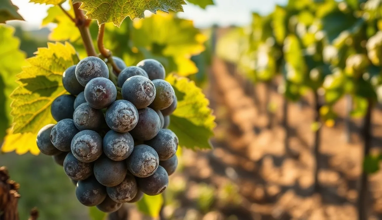
Powdery mildew (Erysiphe necator, formerly Uncinula necator) is the most economically damaging fungal disease of grapevines worldwide, and it's also one of the easiest to photograph [1].
On young leaves, you'll see a dusty white-to-gray coating that sits on either the upper or lower surface. It looks like someone blew flour across the leaf. Unlike downy mildew, the growth stays on top of the tissue instead of inside it. Rub it with your finger and it smears off. That's your fast field test.
On berries, an early infection (pre-veraison) leaves a netlike russeting or cracking as the berry keeps growing but the infected skin doesn't. Berries infected before they reach 8 to 10 mm across are the most vulnerable [1]. Late-season berry infections show the same white coating and often open the door to Botrytis.
On canes and rachis, look for bleached or rusty-brown patches with a diffuse gray dusting. Those wood sites are where the fungus overwinters, either as chasmothecia (dark brown specks you'll need magnification to see) or as mycelium tucked in dormant buds.
The photo mistake people make is confusing mildew with sulfur or kaolin residue. Spray residue is uniform and follows the spray pattern. Mildew is patchy and follows leaf architecture, favoring the shaded interior of the canopy. Shoot in natural light, skip the midday direct sun, and photograph both leaf surfaces.
How do you identify downy mildew vs. powdery mildew in photos?
This is the most common misidentification in the field, and getting it wrong means you reach for the wrong chemistry.
Downy mildew (Plasmopara viticola) has a very different visual signature from powdery mildew. Both produce a white coating, which is where the confusion starts.
Here's what separates them in a photo:
| Feature | Powdery mildew | Downy mildew |
|---|---|---|
| Where white growth appears | Upper OR lower leaf surface | Almost always lower leaf surface only |
| Upper leaf symptom | White coating directly | Yellow-green oily "oil spots" |
| Texture of white growth | Powdery, rubs off easily | Fluffy, cottony, denser |
| Affected plant parts | Leaves, shoots, clusters | Leaves, shoots, inflorescences |
| Climate association | Hot, dry weather | Wet, humid conditions after rain |
| Geographic range | Almost all wine regions | Rarer in very dry regions (Central Valley CA) |
For downy mildew, flip the leaf over. On the underside you'll find a white cottony sporulation directly below the yellow oil spot on top. That yellow-green angular patch on the upper surface, boxed in by leaf veins, is the "oil spot," and it's diagnostic. Cornell's grape IPM program keeps sharp reference images of both diseases [2].
Downy mildew can also hit clusters, turning them brown and collapsing them in a symptom some European literature calls "brown rot of the bunch" or "grey rot." Don't confuse that with Botrytis.
What does Botrytis bunch rot look like in grape clusters?
Botrytis cinerea is the gray mold you know from strawberries, but in grapes it changes character with the season and the variety.
Early on, Botrytis blights young shoots and inflorescences. Tissue goes brown and water-soaked, then turns gray and collapses. That gray comes from masses of conidia (spores) on the surface. Growers call this stage blossom blight.
At harvest, Botrytis is the rot you photograph in tight-clustered varieties like Pinot Noir and Chardonnay. A few berries turn water-soaked, then brown, then collapse under a gray fluffy coating. It moves berry to berry through contact. High humidity and any wound (insect, mechanical, or another fungus) speeds it up fast.
At the gray-mold stage, Botrytis is unmistakable in a photo. The texture is soft and powdery-gray, nothing like the brown leathery look of Aspergillus or the hard mummies of black rot. If intact berries sit right next to gray-molded ones, that inward spread from contact points is a strong tell.
Noble rot (the Botrytis you want for Sauternes-style wines) looks close, but it lands on ripe, dry-skinned berries during a specific weather cycle: morning fog that burns off by afternoon. Those berries shrivel and turn brown-gold with no gray surface. See gray fuzz? You have bunch rot, not noble rot.
WSU's Viticulture and Enology program publishes extension guides that walk through Botrytis timing, with photos, for the Pacific Northwest [3].
How do you identify Eutypa dieback in the field?
Eutypa dieback (Eutypa lata) is a wood disease. That means the canopy symptoms you photograph show up long after the infection settled into a pruning wound.
The spring symptom people photograph most is stunted shoot growth. One or two shoots off an arm or spur come out much shorter than their neighbors, with small, cupped, distorted leaves and dead margins. The leaves look pale, chlorotic, and tattered. That's the Eutypa stunting symptom, and it comes from toxins the fungus makes in the infected wood, not from the fungus touching the leaves.
The diagnostic photo is a cross-section of the infected arm or cordon. Cut into the wood with a handsaw and shoot the cut face. You'll see a wedge-shaped or fan-shaped area of dark brown-to-gray stained wood (necrotic xylem). That V pointing inward toward the pith is the giveaway for Eutypa and its trunk-disease cousins. It looks different from the brown circular staining of Esca.
Eutypa enters through pruning wounds bigger than about 1 cm across, and the fungus can travel 30 cm or more from that entry point before symptoms appear [4]. So a vine flagging leaf symptoms now may have picked up the infection at a cut made three to seven years ago. UC Davis has done the foundational work on Eutypa and trunk diseases and publishes diagnostic guides online [4].
For your records, shoot both the canopy symptom and the wood cross-section. Date it, note the variety, mark the vine number. That's what lets a PCA judge whether the disease is on the move.
What are the visual signs of Phomopsis cane and leaf spot?
Phomopsis viticola shows up in wet spring weather and photographs cleanly once you know the pattern.
On green shoots, look for dark brown-to-black circular lesions on the first few internodes near the shoot base. They're small (2 to 5 mm), ringed by a yellow halo, and they cluster on internodes 1 through 5, right where you count spurs. A badly infected shoot base goes dark, cracked, and bleached after a few weeks, the classic "basal bleaching."
On leaves, Phomopsis makes angular yellow spots with dark centers, concentrated along the midrib and main veins. That linear, vein-following pattern is a solid photo key for telling Phomopsis apart from downy mildew.
On clusters, Phomopsis rots berries late in the season. Infected berries go brown and shriveled, with black pycnidia (fruiting bodies) dotting the surface. This is the late-season problem that hurts wine quality.
The disease overwinters in infected cane tissue and bark, so any block that carried heavy Phomopsis last year is your top risk this spring. Photograph the pycnidia on dormant wood with your phone's macro setting. They look like tiny black dots embedded in the tissue, and they're the source of your spring inoculum.
How do you recognize Pierce's disease symptoms in photos?
Pierce's disease (PD), caused by the bacterium Xylella fastidiosa, is fatal to most Vitis vinifera varieties in regions where the glassy-winged sharpshooter (GWSS) is established [5]. Catching it early from photos matters because there's no cure. Infected vines get removed to slow the spread.
The most photographed PD symptom is leaf scorch. The leaf margin turns yellow-green, then tan-to-brown, working inward. The dead tissue is dry and crispy, and the pattern often leaves a yellow or green band between the dead margin and the healthy center. Heat and drought stress look uniform across the leaf. PD scorch is marginal.
A second tell: irregular green islands sitting inside otherwise dead leaf tissue. That mosaic of living and dead tissue on one leaf points hard at PD.
Late summer, look for "matchstick canes," where the cane fails to lignify in patches and leaves green sections along an otherwise brown cane. Bend the green bark and the wood under it is still soft.
Petioles often stay attached to the cane after the leaves drop in fall, because the abscission layer never forms right. Photograph canes in late October or November. Healthy vines have clean nodes where leaves dropped. PD-infected canes still have dried petioles sticking out.
If you suspect PD, call your county agricultural commissioner. In California, Pierce's disease is reportable in counties with GWSS, and the California Department of Food and Agriculture (CDFA) runs active monitoring programs [5].
What does black rot look like on grapes and leaves?
Black rot (Guignardia bidwellii) does its worst damage in the humid eastern US. In California it's rare, showing up only in high-humidity coastal or inland-valley pockets.
On leaves, the first sign is a reddish-brown circular spot with a dark border, usually 5 to 15 mm across, with a tan or light-brown center. Look inside the spot with a hand lens or your phone's macro setting and you'll see tiny black pycnidia arranged in a ring. That ring of black dots inside the lesion is diagnostic for black rot.
On berries, black rot is dramatic. Berries go from green to brown, then shrivel fast into hard, black, wrinkled mummies that usually cling to the cluster. Those mummies are packed with fungal fruiting bodies and become the main inoculum for next season. Photograph them. They're easy to catch during dormant scouting.
Black rot infection periods track temperature and how long the tissue stays wet. Cornell's NEWA disease prediction models pull hourly weather data to forecast infection risk [2]. If you farm an eastern region, run those models before every significant rain.
What do crown gall and fanleaf look like, and how do photos help?
Crown gall and fanleaf degeneration both come from pathogens that, once established, live in or on the vine for good. Photos document their presence, but they can't replace a lab test.
Crown gall (Agrobacterium vitis) throws up irregular, warty, tumor-like galls at or below the soil line, on the trunk, or at bud unions. Young galls are soft and white-to-cream. Older ones are hard, brown, corky, and can reach baseball size or bigger. They show up best after winter pruning while the vine is dormant. The galls choke off water and nutrient flow, so the canopy collapses on one side during the season. Photograph the gall in context: gall, trunk below it, canopy above it, all in one frame if you can manage it.
Grapevine fanleaf virus (GFLV), carried by the dagger nematode (Xiphinema index), produces two looks. The fanleaf type makes distorted, almost flat leaves with irregular margins, exaggerated teeth, and a fan shape instead of the normal lobes. The yellow mosaic type paints the whole leaf, or patches of it, bright yellow. Both forms shorten internodes and throw shoots into a zigzag. Infected vines decline over years. There's no cure. Retraining or replanting on nematode-resistant rootstocks is the long game [6].
Fanleaf can mimic herbicide injury, especially from hormone-type herbicides like 2,4-D. The photo difference: herbicide injury usually hits only the current season's growth after a spray, often shows a gradient from hardest-hit to least-hit shoots, and lines up with known spray activity nearby.
What tools and apps help identify grapevine diseases from photos?
Here's the honest answer: AI photo-ID tools for plant diseases are getting better fast, but as of 2025 they're not reliable enough to be your only diagnostic source. Studies report 70 to 90 percent accuracy under controlled conditions, and accuracy drops hard on real field photos with mixed symptoms or bad lighting.
The resources that actually earn their keep:
UC IPM, University of California (https://ipm.ucanr.edu): the grape pest management guidelines have disease identification with photos, management timing, and product recommendations built for California conditions [1].
Cornell CALS Viticulture (https://grapesandwine.cals.cornell.edu): disease and pest management pages with extension publications and reference photos for the eastern US [2].
WSU Viticulture and Enology (https://viticulture.cahnrs.wsu.edu): the best fit for the Pacific Northwest, with Botrytis and mildew guides written for cooler, wetter regions [3].
Plant Village (https://plantvillage.psu.edu): a Penn State-backed photo database with crowdsourced identification. Good for a second opinion [10].
If you run a formal spray program, photo documentation is only half the compliance picture. When you need spray dates, PHI intervals, and scouting records in one place, a tool like VitiScribe lets you attach field photos straight to spray records, which pays off when you're trying to reconstruct why you sprayed what you sprayed three months later.
When you're stuck, send physical samples. University plant diagnostic labs (UC Davis, Cornell, WSU, Penn State) confirm visual calls with molecular testing, usually $25 to $75 per sample depending on the lab and the tests you order.
What should a good disease scouting photo include for records?
A phone photo with no context lands in your PCA's inbox and does nothing. Here's what to send instead.
Start with a close-up of the symptom, filling the frame. Then a mid-range shot showing the symptom in place on the vine. Then a block-level shot showing roughly how many vines are hit. Three photos, thirty seconds.
For close-ups, put a known object in the frame for scale. A coin, a glove finger, a pruning shear blade. Phone cameras don't convey scale on their own, and the gap between a 3 mm black rot pycnidium and a 15 mm lesion changes the diagnosis.
For leaves, always capture both surfaces. Flip the leaf and shoot the underside. Downy mildew is nearly invisible from the top. Get the petiole too.
For wood diseases, cut a cross-section and shoot the cut face. Include the outer bark and the inner wood pattern in the same frame when you can.
Metadata is your record. Your phone timestamps every shot automatically, and most phones embed GPS coordinates. Don't edit photos before you send them to an advisor. That original EXIF data is your documentation.
EPA's Worker Protection Standard (WPS) requires employers to keep pesticide application records for at least two years [7]. Scouting photos tied to spray records help show your IPM decisions were evidence-based, which matters during an audit and during any pesticide residue dispute with a buyer. Photos filed by block and date, rather than dumped in your camera roll, are the difference between useful records and a mess.
Which grapevine diseases cause the most economic loss, and what do their damage patterns look like in photos?
National loss data for grapevine diseases is genuinely hard to pin down, because most surveys are regional and variety-specific. The clearest numbers come from a handful of sources.
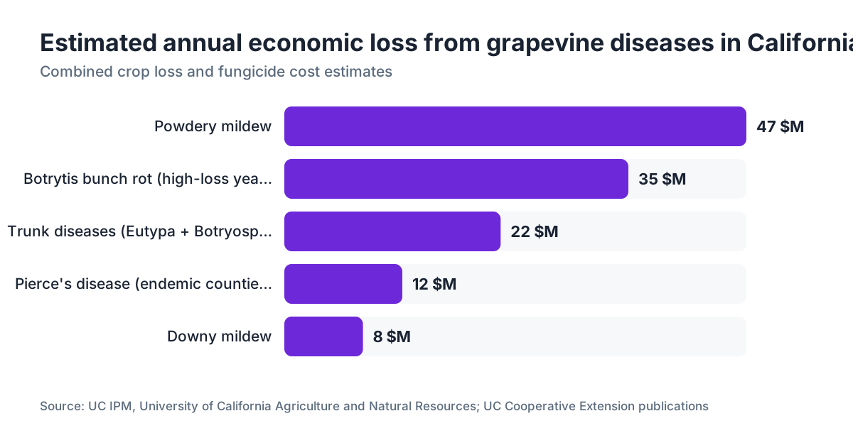
Powdery mildew costs California more than any other grape disease. UC Cooperative Extension surveys put the estimate at $37 million to $57 million a year, fungicide plus crop loss combined [1]. It's the reason sulfur is still the most widely used fungicide in American vineyards.
Botrytis bunch rot runs from 5 percent to 80 percent of the crop in a bad year, depending on variety and harvest timing [8]. Tight-clustered varieties (Pinot Noir, Muscat) in wet years carry the highest risk. Because those losses hit at harvest, they show up in photos as gray rot racing through clusters you can't afford to leave hanging.
Eutypa dieback and other trunk diseases (Esca, Botryosphaeria dieback) together affect an estimated 15 to 50 percent of vines in some older California vineyards [4]. Trunk diseases kill vines a little at a time over years, not in one bad vintage. The real cost is the vine itself plus the three to five years of replanting and training to replace it.
Pierce's disease in Southern California and Texas makes commercial Vitis vinifera production close to impossible in certain counties without resistant varieties or GWSS control [5]. That's less a yield loss than a question of whether the crop is viable at all.
For your own operation, mapping and photographing symptoms across blocks over several years lets you calculate block-level pressure and aim your spray program where it earns its cost. Not glamorous. But it cuts applications you don't need.
How do you document grapevine disease photos for spray record compliance?
Most US spray record rules are set at the state level, and California's are the strictest under the California Department of Pesticide Regulation (CDPR). Growers with more than $5,000 in gross agricultural sales must file pesticide use reports (PURs) that document the pest, the application rate, PHI compliance, and the field location [9].
Photographed disease evidence supports the "pest to be controlled" column of that PUR. An inspector reviewing your records wants to see you had a reason to spray. A dated photo showing 15 percent powdery mildew incidence in Block 4 on May 14 beats "powdery mildew present" scrawled in a logbook.
For WPS compliance, EPA requires that workers and handlers get training, have access to pesticide safety information, and that employers keep specific records available for inspection for two years after the application [7]. The WPS doesn't require scouting photos, but they reinforce the whole documentation picture.
For USDA Organic certification, your certifier asks to see your Organic System Plan (OSP) and records proving you followed it. Scouting records, photos included, show your spray calls came from observed pressure rather than the calendar. That matters when you're explaining why you applied an OMRI-listed copper fungicide on a given date.
For USDA GAP (Good Agricultural Practices) audits, which many grocery-chain buyers require, scouting records and spray logs get reviewed together. Photos attached to spray decisions make the audit faster and cleaner.
Organizing photos is the part most small operations blow. A folder structure by year, block, and date on a shared drive is the floor. If you run a formal spray program, VitiScribe attaches field photos directly to spray records at the application level, which speeds up pulling audit documentation. For larger operations in regions like Paso Robles or the South Coast, where block counts and spray events pile up fast, a structured record system starts paying for itself in the time you save during harvest audits.
Frequently asked questions
What are the most common grapevine diseases I'll see in the vineyard?
The most common are powdery mildew, downy mildew, Botrytis bunch rot, Phomopsis cane and leaf spot, Eutypa dieback, and black rot. Pierce's disease is critical in California, Texas, and the Southeast. Crown gall and fanleaf degeneration are long-term vine health concerns. Which ones matter most depends on your region, climate, and variety.
How can I tell the difference between powdery mildew and downy mildew from a photo?
Powdery mildew shows white powdery growth on the upper or lower leaf surface that rubs off easily. Downy mildew shows a yellow-green oil spot on the upper surface and a fluffy white cottony growth on the underside directly beneath that spot. Flip the leaf over: if the white growth sits only on the underside under a yellow patch, it's downy mildew.
Can I use an app to identify grapevine diseases from photos?
AI plant disease apps give you a starting point but aren't reliable enough for a definitive diagnosis. Accuracy in controlled studies runs 70 to 90 percent, and field conditions produce worse results. Use apps to narrow your suspect list, then confirm with a UC Davis, Cornell, or WSU extension resource or a plant diagnostic lab. For anything affecting a spray decision, spend the $25 to $75 for lab confirmation.
What does Pierce's disease look like, and when should I be worried?
Pierce's disease shows leaf scorch with a yellow-green band between living and dead tissue, green islands on otherwise dead leaves, failed cane lignification (soft green patches on brown canes), and dried petioles staying attached after leaf drop in fall. If you're in a glassy-winged sharpshooter region of California and see these symptoms, call your county agricultural commissioner. There's no cure and it's reportable in some counties.
What does Eutypa dieback look like on a grapevine?
In the canopy you'll see stunted shoots with small, cupped, chlorotic leaves and brown necrotic margins, usually on one arm or spur while the rest of the vine looks fine. Cut the affected arm and you'll find a wedge-shaped dark brown stain in the wood cross-section. That V-shaped internal discoloration is diagnostic for Eutypa and related trunk diseases.
How do I photograph grapevine disease symptoms for a PCA or extension advisor?
Take three photos: a close-up of the symptom with a coin or finger for scale, a mid-range shot showing the symptom in context on the vine, and a block-level shot showing the spread pattern. For leaves, photograph both surfaces. For wood diseases, cut and photograph the cross-section. Don't edit photos before sending; the original timestamp and GPS metadata is your record.
What does Botrytis bunch rot look like at harvest?
Infected berries appear water-soaked and turn brown, then collapse under a soft gray fluffy coating that's the fungal sporulation. The rot spreads berry to berry through contact. It looks different from black rot (which mummifies hard) and Aspergillus (drier and darker). Tight-clustered varieties in wet years carry the highest risk. Photograph the cluster from the side to show the spread pattern.
Does grapevine disease scouting need to be in my spray records?
California growers above $5,000 in gross sales must document the pest being controlled in their pesticide use reports. Dated scouting photos support that documentation and show an inspector your spray decisions were evidence-based. EPA Worker Protection Standard records must be kept for two years. For organic certification, scouting records show applications were triggered by observed pressure, not a calendar schedule.
What do crown gall symptoms look like in photos?
Crown gall produces irregular, warty tumors at or below the soil line, on the trunk, or at bud unions. Young galls are soft and whitish; older ones are hard, brown, and corky and can reach baseball size or larger. They show best after dormant pruning. To document crown gall, photograph the gall in context, showing the gall, the trunk below it, and the canopy above it.
What is the economic impact of powdery mildew on grapevines?
UC Cooperative Extension surveys estimate powdery mildew causes $37 million to $57 million in annual losses in California alone, combining fungicide costs and crop losses. It's the leading cause of annual disease loss in California vineyards and the main reason sulfur remains the most widely used fungicide in American wine grape production.
How does black rot look different from Botrytis bunch rot in photos?
Black rot mummifies berries into hard, shriveled, black raisins that usually stay attached to the cluster. Botrytis produces soft, wet, collapsing berries with a gray fluffy coating. Black rot lesions on leaves carry a ring of tiny black pycnidia inside the lesion. Botrytis is more common at harvest in humid conditions; black rot is a spring and early summer disease in wet eastern climates.
Where can I find free online reference photos for grapevine disease identification?
UC IPM's grape pest management guidelines, Cornell's CALS grape disease pages, and WSU's Viticulture and Enology extension resources all publish free reference photos. For eastern US conditions, Cornell's NEWA disease forecasting system is also worth using. Penn State's Plant Village database has crowdsourced reference photos. For lab confirmation, state plant diagnostic labs typically charge $25 to $75 per sample.
Sources
- UC IPM, University of California Agriculture and Natural Resources, Grape Pest Management Guidelines: Powdery mildew causes an estimated $37-57 million in annual losses in California vineyards; berries infected before 8-10 mm diameter are most vulnerable to skin cracking
- Cornell University CALS, New York State IPM Program, Grape Disease and Pest Management: Cornell's NEWA grape disease forecasting models use hourly weather data to predict black rot and downy mildew infection periods; reference photos available for eastern US conditions
- UC Davis Department of Plant Pathology, Trunk Disease Research: Eutypa lata infects through pruning wounds larger than approximately 1 cm; fungus can travel 30 cm or more from infection point before symptoms appear; trunk diseases affect 15-50% of vines in some older California vineyards
- California Department of Food and Agriculture, Pierce's Disease Control Program: Pierce's disease caused by Xylella fastidiosa is reportable in California counties with established glassy-winged sharpshooter populations; fatal to most Vitis vinifera varieties with no cure
- UC Davis Foundation Plant Services, Grapevine Virus Diseases: Grapevine fanleaf virus (GFLV) is transmitted by the dagger nematode Xiphinema index; no cure exists; management involves replanting on nematode-resistant rootstocks
- EPA, Worker Protection Standard for Agricultural Pesticides: EPA Worker Protection Standard requires employers to keep pesticide application records for at least two years after the application and make them available for inspection
- UC Cooperative Extension, Botrytis Bunch Rot of Grape, Publication 7120: Botrytis bunch rot can cause losses ranging from 5% to 80% of the crop in severely affected years, depending on variety and harvest timing
- California Department of Pesticide Regulation, Pesticide Use Reporting: California growers with more than $5,000 in gross agricultural sales must keep pesticide use reports documenting pest, application rate, PHI compliance, and field location
- Penn State Extension, Plant Village Disease Identification Database: Plant Village provides a crowdsourced photo database for plant disease identification, backed by Penn State University
Last updated 2026-07-09