Grapevine diseases pictures: what you're actually looking at in the field
TL;DR
- The grapevine diseases you'll photograph most in the field are powdery mildew, downy mildew, Botrytis bunch rot, black rot, and Eutypa dieback.
- Each has a distinct visual signature on leaves, canes, or clusters.
- Correct identification drives correct treatment.
- Matching your photo to the right pathogen is step one before you open any spray record or reach for a fungicide.
Why do grapevine disease pictures matter for diagnosis?
A photo is not a diagnosis. That's the honest starting point. But a good close-up of a diseased grapevine leaf is the fastest way to get a second opinion from your farm advisor, your extension plant pathologist, or an AI-assisted ID tool, and it creates a timestamped field record you can attach to your spray log.
Grapevine diseases fool you early. Powdery mildew and downy mildew are both white, but they grow on different leaf surfaces and come from completely different organisms. Black rot lesions and Phomopsis leaf spots both make circular marks on leaves, but black rot turns berries into hard, mummified pebbles. Get the pathogen wrong and you use the wrong chemistry at the wrong timing. That costs money, and worse, it leaves the disease running.
University extension programs at UC Davis [1], Cornell [2], and Washington State University [3] all publish photo-based disease keys for exactly this reason. Those resources are free, updated by plant pathologists, and calibrated to real field conditions in their regions. Cross-check any photo there first.
One habit pays off constantly: photograph both the top and underside of the same leaf whenever you see a symptom. Many fungal pathogens (downy mildew especially) sporulate almost only on the lower surface. A top-side-only photo can be genuinely inconclusive.
What does powdery mildew look like on grapevines?
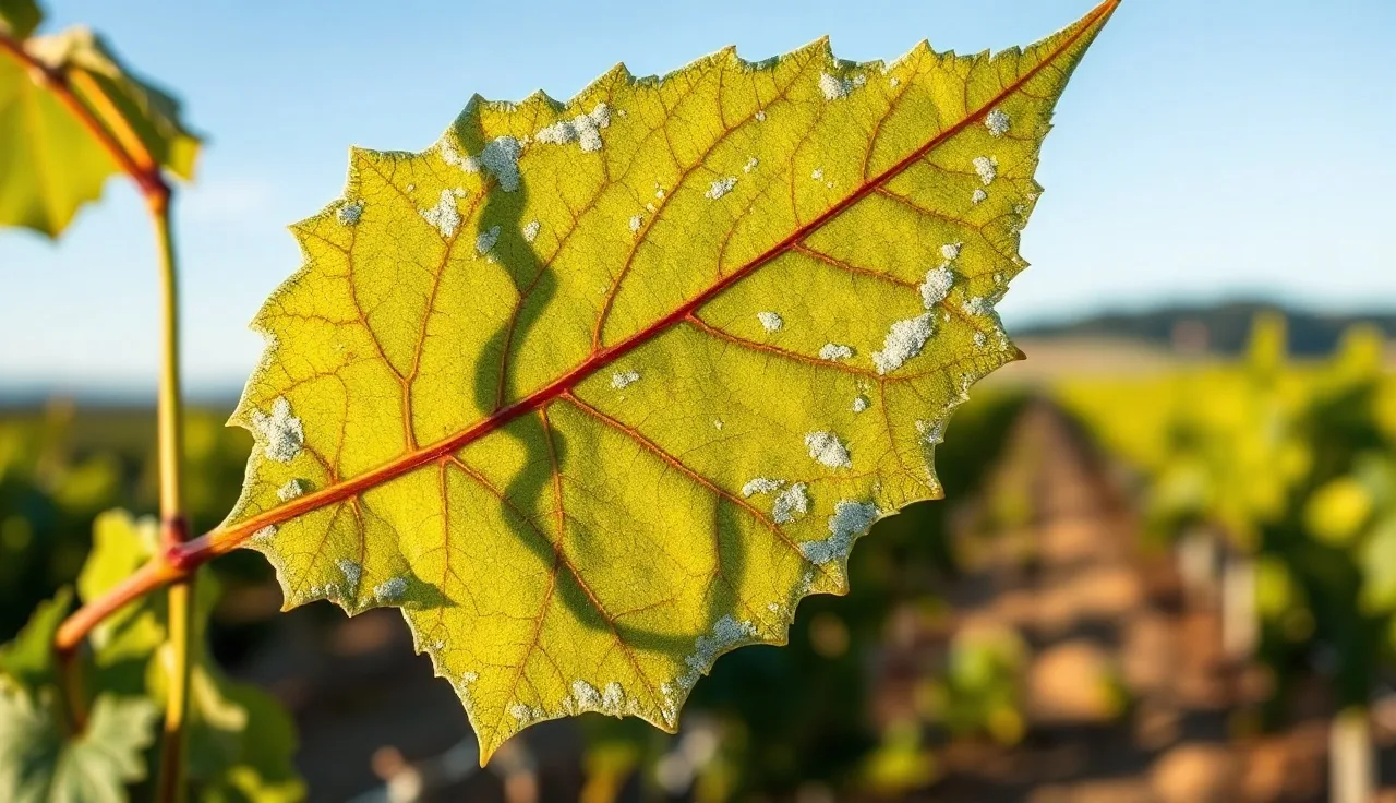
Powdery mildew (Erysiphe necator, formerly Uncinula necator) is the highest-cost grapevine fungal disease in most North American wine regions [1]. The visual signature is hard to miss once you've seen it. A powdery white or gray flour-like coating grows on the upper surface of leaves, young shoot tissue, and developing berries. That coating is fungal mycelium and conidia growing directly on the plant surface. Almost every other grape disease hides inside the tissue. This one grows on top of it.
On leaves, look for discrete white patches, often starting near the midrib or in dead-air zones with poor airflow. Infected tissue under the patch turns yellow or brown as the season runs on. On berries, infection before bloom or in the early post-bloom window (roughly 3 to 6 weeks after capfall) does the most damage. Infected young berries stop expanding and split, opening entry points for Botrytis. Later infections on mature berries carry the white coating but do less structural harm.
Cane infection shows at harvest as reddish-brown or bleached patches on one-year wood. Document this in late season. Heavily colonized canes are a primary inoculum source the following spring.
Powdery mildew is one of the easier diseases to photograph clearly. The white coating pops against green tissue, and a standard phone photo captures it fine. UC Davis Viticulture and Enology publishes a detailed symptom photo set [1].
What does downy mildew look like on grapevine leaves?
Downy mildew (Plasmopara viticola) is a completely different organism from powdery mildew, an oomycete rather than a true fungus, and it needs free water to spread. Leaf symptoms start as oily, angular yellow spots on the upper surface, often called 'oil spots.' Leaf veins bound the spots, which gives them that angular polygon shape that's a reliable diagnostic clue.
Flip the leaf. Look at the spot from below during high humidity or early morning, and you'll see a white cottony sporulation layer. That's the definitive confirmation. No other common grape disease makes that exact combination: yellow oil spot on top, white fuzz underneath.
In California's dry summers, downy mildew is less of a year-round threat than it is in the humid East, but irrigation practices and foggy coastal conditions create enough leaf wetness to trigger infections [1]. In the Finger Lakes, Ohio, and Virginia, it can strip a canopy without a solid spray program.
Berry infection (called 'grey rot of the cluster' in early season, distinct from Botrytis) makes brown, leathery berries that never develop right. The infected cluster stem can turn brown and collapse too, a symptom called 'brown rot of the rachis.' Cornell's disease ID resources include close-up photos of every stage [2].
Estimate lesion density per leaf (light, moderate, heavy) when you photograph. That estimate feeds spray timing decisions directly and makes a useful field note on your pesticide application record.
How do you identify Botrytis bunch rot in photos?
Botrytis cinerea is a generalist fungal pathogen that hits nearly every crop, but it's especially damaging in grapes because of how it moves through a cluster. The classic late-season visual is a gray-brown fuzzy mass of sporulation on shriveled berries. The gray comes from the mass of conidia. Disturb an infected cluster and you'll often see a puff of gray dust lift off.
Earlier in the season, Botrytis shows as flower infection (called 'blossom blight'), where flower caps and stamens don't fall cleanly and infected tissue browns. On young berries, a tan or brown water-soaked lesion at a point of injury (hail damage, insect feeding, a powdery mildew split) is the common entry. The fungus does not push through sound, intact berry skin easily. That's why cluster architecture, spray timing, and any prior berry damage tie so tightly to Botrytis risk.
On leaves, Botrytis makes irregular brown lesions, sometimes with concentric zonation, but leaf infection is rarely the economic concern. The cluster is where the damage happens.
In wine grape production, the split between early Botrytis (before 50% berry infestation) and 'noble rot' used on purpose for sweet wine comes down mostly to humidity and harvest timing. Same pathogen. A sauternes-style infected berry looks grey, shriveled, and concentrated. High-humidity rot looks wet and mushy.
WSU's extension program publishes thresholds and timing recommendations for Botrytis management with photo references tied to its regional conditions [3].
What does black rot look like on leaves, berries, and canes?
Black rot (Guignardia bidwellii) is the dominant fungal disease in humid Eastern US grape production and causes total cluster loss in bad years without a spray program [2]. The leaf symptom starts as small pale green or yellow circular spots that expand to about 0.25 to 0.5 inches across, with a brown center and a yellow halo. Inside the brown necrotic zone sit tiny black pycnidia (spore-producing structures) arranged in concentric rings, sometimes visible with the naked eye and definitely visible with a hand lens. That's the clincher: tiny black dots inside the lesion.
The berry symptom is the most dramatic visual in grapevine disease photography. Infected berries turn brown and soft briefly, then desiccate fast into hard, wrinkled, blue-black 'mummies' that stay attached to the cluster. These mummies are the primary overwintering inoculum for the next season, so their presence in the canopy at harvest tells you exactly what your spray timing missed.
Cane lesions from black rot run elongated, dark brown to black, often with the same tiny pycnidia on the surface. The critical berry infection window runs from just before bloom through about 4 weeks post-capfall, which overlaps heavily with the powdery mildew window. Programs in humid regions are often built to hit both pathogens at once during this stretch.
How do you recognize Eutypa dieback and other trunk diseases?
Trunk diseases are the slow-moving catastrophe of viticulture. They don't kill vines overnight. They accumulate over years and eventually force retraining or replanting. The main trunk diseases in North American vineyards are Eutypa dieback (Eutypa lata), Botryosphaeria dieback (several Botryosphaeriaceae species), the Esca/Petri disease complex, and Phomopsis cane and leaf spot.
Eutypa dieback visual signature: in spring, one or more arms of an infected vine push short, stunted, chlorotic (yellow) shoots with necrotic leaf margins while adjacent arms look healthy. That uneven spring growth is the most reliable field indicator. Cut into the affected wood and you'll see a wedge-shaped brown necrotic sector in the cross-section. Photograph that internal wood symptom too, more than the exterior, because the external cankers can be subtle.
Esca complex makes what's called 'tiger stripe' symptoms on leaves: interveinal chlorosis and necrosis that creates alternating green and brown stripes along the leaf veins. In severe cases the whole vine collapses suddenly in mid-summer, a symptom called 'apoplexy.' Tiger-stripe leaf photos are distinctive and easy to cross-reference.
Phomopsis cane and leaf spot (Phomopsis viticola) makes elongated black lesions with yellow halos on the basal internodes of young canes in early spring, plus small circular spots with yellow halos on leaves. Berry infection from Phomopsis produces a soft brown rot called 'dead arm rot' in some older literature.
All trunk disease management hinges on protecting pruning wounds, so the timing and method of your pruning cuts are the primary intervention. UC Davis has published extensively on trunk disease epidemiology and wound protection timing [1].
What are the main grapevine virus diseases and how do they look?
Viruses are a different animal from fungal or bacterial diseases because there's no spray to cure them. The economic decision is whether to pull the vine and replant with certified clean material. So accurate visual identification matters enormously.
Grapevine leafroll disease is the most widespread grapevine virus problem on earth. In red varieties the visual is iconic: leaves roll downward along their margins in late summer and fall and turn red or purple between the veins while the veins stay green. In white varieties the interveinal yellowing happens without the red color, which makes it harder to spot. Cornell publishes virus ID guides with photos calibrated to Eastern US varieties [2].
Grapevine fanleaf virus causes severely distorted, asymmetric leaves with deep serrations (hence 'fanleaf'), yellow mosaic patterns, and in some strains, yellow vein banding. Affected vines set fruit poorly and grow short internodes and fasciation (flattened, fused shoots). The dagger nematode (Xiphinema index) spreads this virus in soil, which is why replanting without soil fumigation or a long fallow period often just reinfects the new vines.
Red blotch disease, caused by Grapevine red blotch virus (GRBV), was only formally characterized in 2012 [4]. Leaf symptoms mimic leafroll: red blotching and marginal reddening in red varieties. The distinction matters because the vector and the management differ. Red blotch spreads via the three-cornered alfalfa treehopper (Spissistilus festinus) and has a different epidemiology. Serological testing (ELISA) or PCR confirmation is necessary for a definitive virus diagnosis. Photos alone can't separate leafroll from red blotch reliably.
What do common insect grapevine pests look like, and how do they interact with disease?
Insect pests and diseases often land in the same search because insect damage opens entry points for fungal pathogens. The connection is real and worth understanding.
Grape berry moth (Paralobesia viteana) larvae are small greenish caterpillars that feed inside berries, leaving entry wounds that Botrytis exploits immediately. Externally you see webbing holding berries together plus berry shrivel or discoloration at the feeding site. Frass (insect droppings) inside the cluster is the diagnostic sign.
Grape leafhoppers (Erythroneura species) cause stippling on the upper leaf surface, a fine white or yellow speckling from feeding. Heavy populations strip leaf area and can slow fruit ripening. The insects themselves run tiny (1-3mm) and jump when disturbed, one reliable field ID cue.
Grapevine phylloxera (Daktulosphaira vitifoliae) is the most historically significant grape pest worldwide. The foliar form makes galls on leaf undersides in American species and some hybrids. The root form, which is the devastating one on Vitis vinifera, stays invisible without digging. If you see leaf galls, photograph the gall and the insect inside it if you can crack one open.
Mealybugs (mainly Planococcus ficus and Pseudococcus species in California) are vectors of several grapevine leafroll-associated viruses. The pest and the virus disease are photographically linked. Cottony white waxy masses on canes and under bark are the mealybug sign. Their economic damage covers both direct feeding and leafroll transmission.
Under EPA Worker Protection Standard compliance [5], any worker scouting for pests and collecting samples is doing an agricultural activity in a treated area, so training and personal protective equipment requirements apply based on your pesticide application records. This compliance point is easy to miss during scouting.
What do bacterial grapevine diseases look like in photos?
Bacterial diseases show up less often than fungal ones in most regions, but they cause distinctive symptoms that read differently from fungal infection.
Pierce's Disease (caused by Xylella fastidiosa, spread by glassy-winged sharpshooter and other leafhopper vectors) produces a symptom called 'leaf scorch': irregular brown burning around leaf margins that advances inward, with a yellow or red transition zone between the dead margin and green tissue. A hallmark sign is that the dead margin tissue often falls away, leaving a ragged leaf edge. Infected fruit clusters partially desiccate. Pierce's Disease is lethal to Vitis vinifera and there is no cure. It's a mandatory reporting disease in California [6].
Crown gall (Agrobacterium vitis) produces rough, corky tumor-like galls at the crown (base of vine), on roots, or at graft unions. The galls start small, pale, and spongy, then harden and darken with age. Freeze damage sharply increases crown gall expression because the bacteria colonize wound sites. Photographing galls at the soil line means moving mulch or soil away from the vine base first.
Grape bacterial blight (Xanthomonas campestris pv. viticola) is less common in the US than in some other grape-growing regions but can cause angular leaf spots that look like downy mildew without the underside sporulation, plus cane necrosis and cluster blight. Lab confirmation matters for bacterial diseases because visual ID alone is often not enough.
How do you build a useful grapevine disease photo record for compliance?
A photo on your phone helps only if you can find it in six months and connect it to the spray decision it triggered. That's the gap most vineyard managers hit.
A good field photo record has four parts: date and GPS location (most phone cameras embed this automatically, so check your camera settings), the specific block and row, what you saw (symptom description in plain language), and what action followed. If the photo triggered a fungicide application, the record connects to the spray entry in your pesticide application log. That connection matters for regulatory audits and for your own season review.
Some operations are moving to digital field tools that attach a photo directly to a scouting event or spray record entry. VitiScribe, for example, links field observations to application records so the photo lives in the same place as the compliance paperwork, which simplifies end-of-season auditing.
For farms under the EPA Worker Protection Standard [5] and state pesticide reporting requirements, a documented basis for each spray application, including the pest or disease observed, is increasingly expected. Calendar spraying without documentation is fading as state agencies tighten scrutiny.
For a broader look at how vineyard field operations connect to compliance workflows, see our overview at vineyard.
Store photos in a folder structure by year, block, and date. Back them up off-device. A disease photo archive from 2019 is sometimes exactly what you need when the same problem returns in 2025 in the same block.
Which grapevine diseases cause the most economic loss?
Economic loss from grapevine diseases swings hard by region and season, but a few numbers help calibrate where to focus.
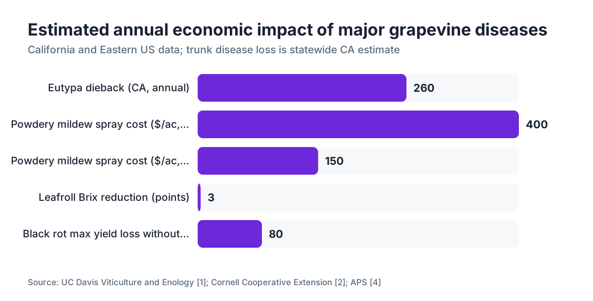
Powdery mildew ranks as the highest-cost grapevine disease in California, with fungicide programs for large operations running $150 to $400 per acre per season depending on chemistry and application count [1]. Nationally, losses from all grape diseases have been estimated at over $1 billion annually in a frequently cited assessment, though that figure aggregates across many pathogens and production systems.
Botrytis bunch rot causes crop loss ranging from a few percent in dry years with open canopies to total cluster loss in high humidity after hail or rain during ripening. UC Davis Cooperative Extension has documented 10-40% yield losses in affected blocks in some California seasons.
Trunk diseases are the sleeper cost. UC Davis researchers found in a survey of California vineyards that Eutypa dieback alone caused estimated annual losses of $260 million statewide [1]. That figure rolls up reduced yield, reduced vine longevity, and replanting costs.
Leafroll virus cuts soluble solids (Brix) at harvest by 1-3 points in infected vines and delays maturity, which in premium wine production hits fruit quality and price directly [4]. The math on pulling and replanting a leafroll-infected block depends heavily on land value, varietal demand, and how far infection has spread through the block.
| Disease | Primary Loss Type | Estimated CA Annual Loss | Key Citation |
|---|---|---|---|
| Powdery mildew | Yield, quality, spray cost | Major (highest spray cost) | UC Davis [1] |
| Eutypa dieback | Vine longevity, yield | ~$260 million | UC Davis [1] |
| Botrytis bunch rot | Direct yield loss | 10-40% in affected blocks | UC Davis [1] |
| Leafroll virus | Quality, maturity delay | 1-3 Brix reduction | APS/Cornell [4] |
| Pierce's Disease | Vine death | Localized, expanding | CDFA [6] |
| Black rot | Yield (Eastern US) | Up to 80% without sprays | Cornell [2] |
Where are the best free photo resources for grapevine disease identification?
The three university extension programs most referenced by professional viticulturists all have freely accessible disease photo libraries.
UC Davis Viticulture and Enology (viticulture.ucdavis.edu) maintains disease fact sheets with photo sets. Its resources are strongest for California conditions: powdery mildew, trunk diseases, Pierce's Disease.
Cornell University's NYS IPM Program and its collaborative 'Grape Disease Control' publication carry photo libraries well-calibrated to Eastern US humid conditions: black rot, downy mildew, Botrytis, leafroll. Cornell Viticulture and Enology distributes these through its cooperative extension network [2].
Washington State University Extension (extension.wsu.edu) has disease and pest identification resources for the Pacific Northwest, including Botrytis timing thresholds and trunk disease management relevant to irrigated Vitis vinifera production in drier climates [3].
The American Phytopathological Society (APS) publishes the 'Compendium of Grape Diseases, Disorders, and Pests,' the peer-reviewed reference standard. It's not free (university library access or purchase), but it's the most thorough single reference available, with high-quality photos.
Beyond extension resources, the UC IPM Online pest management guidelines for grapes [7] include field photos with life-stage information for both diseases and insect pests together, which helps precisely because so many field observations involve both in the same cluster or leaf.
Frequently asked questions
What does powdery mildew look like on grapevine berries?
Infected young berries show a white or gray flour-like coating on the surface. Berries infected before or just after bloom stop expanding and may split or crack. Later in the season, infected berries may carry the white coating but hold their shape. Under the coating, the skin often looks discolored and feels rough. The powdery coating tells it apart from Botrytis, which is gray-brown and fuzzy.
How can I tell downy mildew from powdery mildew in photos?
Powdery mildew shows white powder on the upper leaf surface. Downy mildew shows oily yellow spots on the upper surface and white downy sporulation on the underside of the same spot. Photograph only one surface and you can misidentify both. Always capture both. Powdery mildew is a true fungus; downy mildew is an oomycete and needs free water to spread.
What do black rot mummies look like and why do they matter?
Black rot mummies are desiccated, hard, wrinkled, blue-black berries that stay attached to the cluster after all others drop. They photograph easily because they contrast sharply against other material. They matter because each mummy carries overwintering pycnidia that release spores the following spring, which makes mummy removal or burial an important cultural practice in humid regions.
How do I photograph grapevine leaf symptoms correctly for disease identification?
Photograph the same leaf from both surfaces. Get as close as your phone can focus without blurring, ideally 6-8 inches from the leaf. Avoid direct harsh sunlight, which washes out texture. Capture multiple lesion stages if they're present: early, mid, and advanced. Include a coin or finger for scale. Shoot in natural morning light when you can. Always note the variety, block, and date.
What does Pierce's Disease look like on grapevine leaves?
Pierce's Disease causes 'leaf scorch': brown, irregular burning around leaf margins that advances inward, with a yellow or reddish transition zone between dead and live tissue. The dead margin tissue often falls away, leaving a ragged edge. Infected clusters partially desiccate. These symptoms appear mid-to-late season. It's a mandatory reporting disease in California; contact your county agricultural commissioner if you suspect it.
Can I identify grapevine virus diseases from photos alone?
Not reliably, no. Leafroll and red blotch disease both cause red leaf coloration and margin rolling in fall, and separating them visually is very difficult even for experienced plant pathologists. Fanleaf virus has more distinct symptoms but still benefits from lab confirmation. ELISA or PCR testing on leaf or petiole samples is the accepted diagnostic standard. Use photos to document and prioritize which vines to sample, not to make final calls.
What insect damage on grapevines is most commonly confused with disease?
Leafhopper stippling looks like light fungal spotting from a distance. Phylloxera leaf galls look somewhat like a fungal gall disease. Grape berry moth entry wounds on berries look like early Botrytis before the fungal sporulation appears. The presence of frass, webbing, or live insects helps separate insect damage. A hand lens helps enormously. When in doubt, send a sample to your state extension plant diagnostic lab.
How do trunk diseases show up early enough to photograph and treat?
Eutypa dieback and Esca show early as uneven spring growth: one arm of a vine pushing stunted, chlorotic shoots while adjacent arms look normal. Photograph the whole vine from a few feet back to capture that uneven pattern, then move in for a close-up of the affected shoot tips. On wood, look for internal brown necrosis on a cross-cut of the affected arm. Trunk disease management depends on wound protection at pruning time, so early detection drives next-season strategy.
Do I need to keep grapevine disease photos as part of my pesticide records?
No federal rule currently requires photos in pesticide records, but EPA Worker Protection Standard regulations [5] require records of pesticide applications including the pest or disease being managed. A photo that matches the date and block of each application gives you a documented basis for the spray decision. Some states are moving toward requiring IPM documentation that includes scouting records. Photos are cheap insurance.
What's the best time of day to scout for grapevine disease symptoms?
Early morning is generally best for two reasons: downy mildew sporulation is most visible when humidity is high after overnight leaf wetness, and natural morning light is diffuse enough to show leaf texture without washing out symptoms. Powdery mildew is visible any time. Berry symptoms photograph well in overcast light. Avoid scouting right after rain, because fresh water can obscure symptom color and texture.
What extension resources offer the best grapevine disease photo identification guides?
UC Davis Viticulture and Enology is strongest for California and Western US diseases, especially powdery mildew, trunk diseases, and Pierce's Disease. Cornell's NYS IPM and Viticulture Extension programs cover Eastern US diseases, especially black rot, downy mildew, and Botrytis. WSU Extension covers the Pacific Northwest. For a paid but authoritative reference, the APS Compendium of Grape Diseases is the standard.
How much can grapevine diseases actually cost per acre?
Powdery mildew fungicide programs run roughly $150 to $400 per acre per season in California depending on chemistry and application count [1]. Eutypa dieback losses in California have been estimated at $260 million annually statewide [1]. Leafroll virus infection cuts Brix at harvest by 1-3 points, which affects fruit price in premium wine production. Black rot without a spray program in humid Eastern regions can cause up to 80% crop loss in a bad year [2].
What's the difference between Botrytis and Phomopsis bunch rot in photos?
Botrytis shows gray-brown fuzzy sporulation on affected berries, often visible as a puff of gray dust when the cluster is disturbed. Phomopsis bunch rot (dead arm rot) appears earlier in the season as a soft, light brown rot, often at the berry stem attachment point, without the gray sporulation. Botrytis is far more common. Early Phomopsis infections are often confused with early Botrytis without a lab check.
How do I know if my grapevine photos are good enough for a diagnosis?
A usable diagnostic photo shows the lesion or symptom in focus at close range with enough context to identify the plant part (leaf, berry, cane, trunk). Include both leaf surfaces for any leaf symptom. Your extension plant pathologist or farm advisor can usually give a working diagnosis from a clear close-up paired with variety, region, and date. Blurry, backlit, or single-surface photos routinely come back as 'inconclusive.'
Sources
- Cornell University Cooperative Extension, Viticulture and Enology Program: Black rot can cause total cluster loss in humid Eastern US regions without a spray program; photo ID resources for Eastern US grape diseases
- Washington State University Extension, Grape Pest Management: WSU publishes Botrytis timing thresholds and trunk disease management resources for Pacific Northwest vineyards
- American Phytopathological Society, Grapevine Red Blotch Virus characterization: Grapevine red blotch virus (GRBV) was formally characterized in 2012; leafroll virus infection reduces Brix at harvest by 1-3 points
- EPA Worker Protection Standard, 40 CFR Part 170: Workers scouting or handling samples in treated areas are subject to WPS training and PPE requirements; pesticide application records must document the pest or disease being managed
- California Department of Food and Agriculture, Pierce's Disease Control Program: Pierce's Disease caused by Xylella fastidiosa is a mandatory reporting disease in California; infected Vitis vinifera vines have no cure
- UC IPM Online, Grape Pest Management Guidelines: UC IPM online guidelines include field photos with life-stage information for grape diseases and insect pests
- Cornell University, NYS IPM Program, Grape Disease Control: Cornell NYS IPM publishes photo-based disease identification keys calibrated to Eastern US humid conditions including black rot, downy mildew, and Botrytis
- American Phytopathological Society, Compendium of Grape Diseases, Disorders, and Pests: The APS Compendium is the peer-reviewed reference standard for grapevine disease identification with high-quality photos
Last updated 2026-07-09